Unique Post-Traumatic Presentation of Central Incisors 13 Years Following Initial Trauma
By William Wilson, Jr., DDS, MS
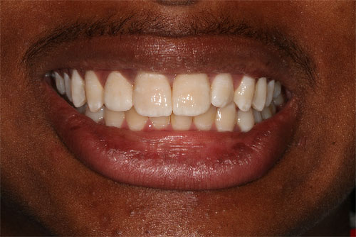
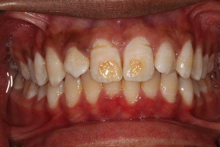
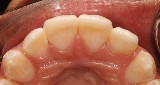
I would like to share an interesting case we had the privilege to treat in our office following a referral from this young man’s general dentist. Darin is 16 years old, and he was unhappy with the appearance of his central incisors. He was attended with supporting and well educated parents inquiring about solutions to improve the esthetics for their son. The maxillary central incisors were reportedly damaged following trauma at the age 3, when the primary teeth were abruptly intruded and made contact with the developing central incisors of the secondary dentition. The enamel of the central incisors did not completely form in the area of trauma, and has the appearance of dentin development beyond the confines of the enamel and onto the surface of the enamel as seen above. This creates a “reverse position” of dentin exterior to the enamel surface in the area of damage, and the resulting unesthetic appearance is certainly noticeable at conversational distance.
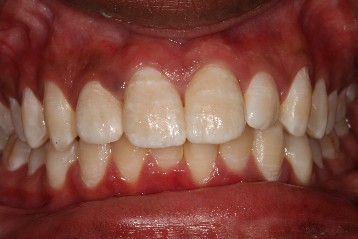
The maxillary and mandibular midlines are coincident, and in harmony with the facial midline, but there is a malformed maxillary right lateral incisor and a mesio-distal space discrepancy between the the two lateral incisors.
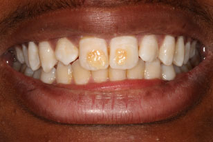
The malformed maxillary right lateral incisor also presents with a “talon cusp”, that will likely have a pulp horn that must be considered for any restorative treatments to be planned. Darin and his parents were informed that ideal treatment would include orthodontics to improve the spacing and allow for symmetry of the lateral incisors, but Darin had waited many years to have this esthetic correction, and was not interested in any further delays. He wanted a more immediate result and was accepting of the concern of the lateral incisors if we could make the discoloration of the central incisors go away. We spent considerable time discussing the clinical presentation and options, and there was an expectation that one lateral incisor would be larger than the other, or continue to have the distal diastema. In addition to the mesio-distal spacing discrepancy, the concerns about symmetry, the dentin on the surface of the central incisors, and the pulp horn, we would also be contending with a hyper-mineralized “banding” in trying to make the ceramics blend with the existing dentition. There was also a groove noted in the cervical 1/3 of the central incisors and concerns about how much enamel would be available for bonding a veneer. Prior to tooth preparation for veneers, we did attempt to remove only the dentin from the surface. The result was unacceptable, and treatment with veneers would yield a more acceptable result.
Problem List:
- Exposed dentin of central incisors creating unacceptable color.
- There will not be enamel to bond at the sites of malformation and the plan may need to change after tooth preparation if 360 degrees of enamel is not present.
- Mesio-distal space discrepancy of maxillary lateral incisors that will create a challenge with symmetry.
- Talon cusp and malformed maxillary right lateral incisor.
- Color science complexities due to the hyper-mineralized “banding”
- Facial groove at the cervical 1/3
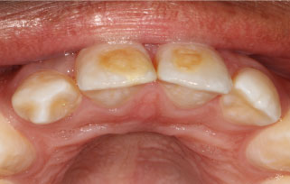
The central incisors were prepared for lithium disilicate veneers. The maxillary right lateral incisor was also prepared, but required reshaping of the tip of the ‘talon cusp’, taking great care to avoid pulpal exposure and required preparation onto the lingual surface. The prep design can be seen in the photos below.
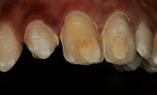
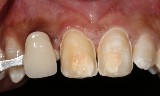

You can also note the defect areas clearly seen in the photo with the polarizing filter applied. The polarizing filtered photographs proved invaluable in the laboratory to aid in color matching.
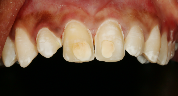
The team at NOVA Prosthodontics achieved an excellent result for this patient, and we thought we matched the color well. After the veneers were bonded to place, Darin and his mother were delighted with the results……until he got home, and then thought the defect was not completely covered, and was “showing through the veneer”.
We later learned, that at home, Darin was inspecting the veneers with a flashlight held behind them attempting to see the defect area. It was not until considerable discussion and review of all the images that the father saw the image with the shade tab and recognized that they were expecting a uniform color, and not the characterized colors, we worked so hard to achieve to make this blend so well. Darin’s father then realized this would not look nearly as good as what we achieved and would actually “stand out”. Everyone then became accepting of the results of treatment. That photo of the shade tab was not something I intended to show, but it allowed the father to see and explain something I was unable to communicate to the patient and family on my own. It truly does take a team!
Treatment completed photos
Comments closed
No comments. Leave first!