Diagnostic Considerations For A Maxillary Implant Supported Prosthesis
The utilization of dental implants as anchorage for dental prostheses has increased considerably in the past decade. Advancements in dental implant treatment has enabled the dental practitioner to treatment plan and deliver complex and challenging prostheses never before thought possible. Dental implant treatment is routinely provided with minimal patient morbidity and with well documented long term clinical success. Unfortunately, the greater than 95 percent osseointegration rate of dental implants has created a false sense of confidence for some dental practitioners in thinking that as long as an implant integrates….anything is possible.
One of the most popular treatment planned dental prostheses is a fixed implant supported prosthesis for an edentulous maxilla. Unfortunately, very often, a fixed maxillary implant supported prosthesis is promised to patients without appropriate diagnosis and treatment planning. Dental implants sometimes are placed without crucial pre-prosthetic surgical procedures and the end result is either an unaesthetic prosthesis or something other than what was promised to the patient.
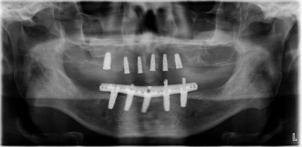
In the panographic image below we can see dental implants that have been placed in both arches. The mandible has been restored while the maxilla awaits final restoration.
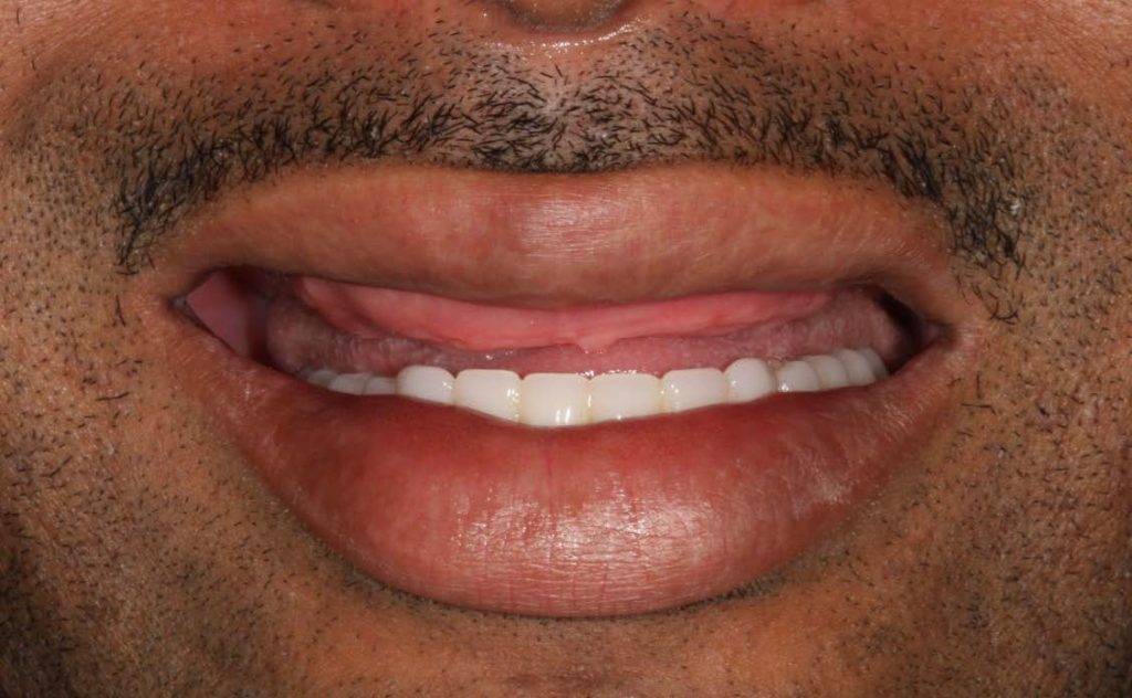
The clinical photograph above is of the same patient whose radiograph is shown. Note the amount of hard
and soft tissue visible in repose in the clinical photograph. Unfortunately, the implants have already been placed, and this scenario demonstrates inadequate pre-prosthetic planning that overlooked the need for a maxillary alveolectomy prior to implant surgery.
Comments closed
No comments. Leave first!